SARS Surface Proteins
SARS Surface Proteins
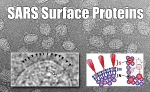
Interpretation of coronavirus virion structure. Conserved structural proteins are drawn as they appear in edge views (left) and axial views (right). Trimeric spikes (S) are shaded in red, membrane proteins (M) are in solid blue, and nucleoproteins (N) are shaded in violet. The dimensions of lattices of S trimers (a 14.0 nm, b 15.0 nm, 100) and RNP molecules (c 6.0 nm, d 7.5 nm, 100) were determined from the reflections shown in Fig. 5A and were consistent with real-space measurements of the same parameters.
Benjamin Neuman


