February 10-14 2003: Distinguished Lectureship Series 2003
Distinguished Lectureship Series 2003
Dr. Michael Moody
10 February 2003 – 14 February 2003
Luckily, merely a superficial grasp was needed to see why the TMV X-ray picture suggested a helix with a turn every 23 A along the helical axis. The rules were, in fact, so simple that Francis considered writing them up under the title, “Fourier Transforms for the Birdwatcher”‘. (Watson, “The Double Helix”.)
All lectures are open to the public. All lectures will be held at noon in the CIMBio ground floor conference room, room 107. Refreshments will be served. For directions to CIMBio see http://cimbio.scripps.edu.
Monday, 10 February; Noon.
I. Basics: Symmetry and Intuitive Fourier Transforms.
- Translational and rotational symmetry.
- Correlations and FTs.
- Basic ideas about 1D and 2D FTs.
- Fundamental FT theorems and application to 1D and 2D FTs.
- Optical diffraction.
- Optical filtering.
Tuesday, 11 February; Noon.
II. Helical Image Analysis: Essentials.
- Helical symmetry.
- FTs of thin helices.
- Flattened and curved helices; selection rules.
- Thick curved helices: FTs and determining helical symmetry.
- Getting 3D structures: general problem; helices.
Wednesday, 12 February; Noon.
III. Helical Image Analysis: Details
- Extracting phase-contrast information from defocused images.
- Digital FTs (general).
- Digital FTs of helices.
- Helix alignment.
- Helix distortion.
Thursday, 13 February; Noon.
AMI Forum
“What is the Future of Molecular-Resolution Electron Microscopy?”
Over four centuries’ quest by microscopists to reveal the most detailed biological structures leaves, as its principal heir, the modern electron microscope and computer image-processing. Invented during the second quarter of the 20th century, electron microscopy’s early development was slowed by the war. Then it made such advances in the third quarter, that it seemed likely that the last quarter would see it become a major technique for finding detailed molecular structures. And yet, despite important developments, it has not lived up to this promise. Is that merely destiny postponed, or must electron microscopy’s role always be limited to an intermediate-resolution structural liaison between molecules and cells?
 |
 |
 |
 |
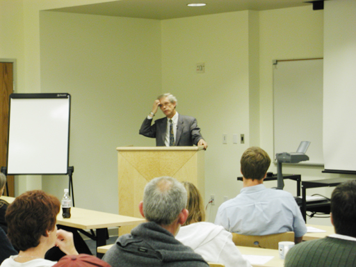 |
Michael M.F. Moody: CV and Publications
| 1958-62 | London University (University College London) Ph.D. in department of J.Z. Young, group of J.D. Robertson. Electron microscopy of fixed/sectioned tissues: membrane origins of vertebrate and some invertebrate photoreceptors. Polarized light sensitivity in Octopus (with J. Parriss). X-ray diffraction of nerve myelin, using swelling to obtain phases, analysed by sampling theorem. |
| 1962-66 | Caltech, Biology Division (groups of A.J. Hodge and of R.S. Edgar) Electron microscopy of negatively-stained T4 phage; image analysis. Symmetry of head and of extended and contracted sheaths. |
| 1966-72 | Rockefeller University, Cell Biology Department of G.E. Palade Helical image analysis of contracted sheaths. Mechanism of T4 sheath contraction established using partially-contracted sheaths. Electron microscopy of tail-tubes shows they are framework for sheath assembly. X-ray scattering of oriented tail-tubes establishes their symmetry (with L. Makowski). |
| 1972-77 | Brookhaven National Laboratory (Biology Division) Solution scattering with neutrons and X-rays: large quaternary structure change found in ATCase (E. coli) by X-ray scattering recorded photographically. Scattering equation for populations of spherical vesicles with heterogeneous radii. |
| 1977-84 | European Molecular Biology Laboratory (Structure Division) ATCase structure change established with electronic recording (laboratory of V. Luzzati, with P. Vachette). Development of apparatus for stopped-flow X-ray scattering using synchrotron radiation sources (with A. Fowler, P. Vachette). Measure speed of ATCase allosteric transition by chemical quenching (laboratory of T. Barman, with H. Kihara).H+-ATPase, membrane-bound c-subunit dissolved in organic solvents studied with NMR (NMR in laboratory of I.D. Campbell, with J. Carver and P. Jones). Evidence for previously suggested hairpin structure. |
| 1984-96 | London University (School of Pharmacy), Chemistry Department (W.A. Gibbons) X-ray solution scattering of ATCase gives R:T proportions related to activity (partly in laboratory of G. Herve and at LURE, also with P. Vachette and J. Fettler): MWC theory inapplicable to CTP inhibition. Kinetics of ATCase allosteric transition by X-ray scattering (with H. Kihara, H. Tsuruta etc.) Review of image analysis of electron micrographs. |
| 1999 | Role of surface curvature in maturational structural transition of phage heads. |

