Analysis of the Drosophila Brain
Analysis of the Drosophila Brain
An Integrated Micro- and Macroarchitectural Analysis of
the Drosophila Brain by Computer-Assisted Serial Section Electron
Microscopy. PLoS Biol 8(10): e1000502. doi:10.1371/journal.pbio.1000502.
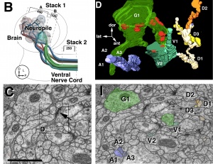
Leginon was used in this study to image total of 500 sections of the
neurophile in one brain hemisphere and 250 sections of the ventral nerve
cord at 4 nm/pixel. (B) and (C) are extracted from Figure 1 of the paper
that show the stack location of the sections imaged and an example region
of the montage acquired on one of the section, respectively. The images
were segmented (I), and combined to reveal the 3D network of in the brain
(D) (Both extracted from Figure 4 of the paper).
Composite Figure from Cardona A, Saalfeld S, Preibisch S, Schmid B, Cheng
A, et al. (2010)


